

BHCC offers state-of-the-art services for ultrasound in Los Angeles & Beverly Hills for accurate medical diagnostics and imaging.
While ultrasound can be used for many purposes, the diagnostic ultrasound at the Beverly Hills Cancer Center is used to detect cancer so we can treat it rapidly, increasing the chances of recovery.
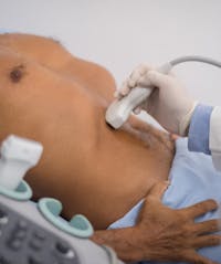
Diagnostic ultrasound, also called sonography or medical sonography, utilizes high frequency sound waves above the range of human hearing to produce images of internal structures. In an ultrasound, the technician uses a small handheld transducer that delivers sound waves into the body. The sound waves return to the transducer and are delivered into a computer, which interprets the waves into a viewable image that provides valuable data concerning internal organs and structures. Because an ultrasound utilizes sound waves instead of radiation, it is an entirely safe procedure.
Ultrasound can be used to help doctors diagnose different types of cancer and evaluate the progression of treatment. Your physician will recommend an ultrasound based on the type of cancer you have. The advantage to ultrasound is it gets pictures of soft tissue diseases that may not show up well on x-rays. It can also be used to tell fluid-filled cysts from solid tumors, which is valuable in the diagnosis of cancer.
Ultrasound services at Beverly Hills Cancer Center include:
- Neck/Thyroid
- Chest
- Renal
- Testicular with Duplex
- Pelvic
- Breast
- Abdomen
- Abdomen Doppler
- Aorta Duplex (AAA)
- Carotid Duplex
- Venous Duplex (Lower and Upper Extremities)
- Arterial Duplex (Lower and Upper Extremities)
- Ankle Brachial Index (ABI)
- Hemodialysis Graft Evaluation
- Vein Mapping
Ultrasound Testing at Beverly Hills Cancer Center
Ultrasound imaging allows doctors to identify a tumor, as well locate the exact area upon which to perform a biopsy. At Beverly Hills Cancer Center, we use the most advanced testing methods and technology in our state-of-the-art facility, including ultrasound imaging.
How Ultrasound Imaging Works
In an ultrasound test, sound waves are sent into the targeted area of the body, and the echo of the sound waves is sent to a computer that creates a real-time image of the systems within the body structure. It is safe and painless, and does not involve the use of radiation.
Preparing for an Ultrasound Imaging Test
Prior to your ultrasound, you will be given directions about what you need to do to prepare. In many cases, all you will need to do is wear comfortable, loose-fitting clothing. Different areas of the body may require more specific pre-test instructions. An ultrasound performed on the abdomen may require you to eat a fat-free meal the evening prior to the test, to avoid food or drink for 12 hours prior to the test, and to drink a quart of water one hour prior to the test so your bladder is full, to help create a better image. Many health insurance plans cover all or part of the cost of ultrasound image tests.
The Ultrasound Test
For the test, you will be asked to remove some (or all) of your clothing, depending upon the area of the body to be viewed. Gel is spread on the skin of the area to be tested. The image is produced with the use of a hand-held device called a transducer, which the technician moves back and forth over the body. Images are displayed on a screen, and these images are saved so they can be reviewed by a radiologist to aid in diagnosis. In some cases, a probe is inserted in the body, such as in cases of suspected prostate cancer or to view the uterus or ovaries in a woman. The test will take only 20 minutes to an hour to perform.
Identifying Tumors with Ultrasound Imaging
The ultrasound imaging test is used to identify tumor growth in various areas of the body. Once an area is identified, a biopsy can be performed to test the tissue for cancer cells. This is termed “image-guided” biopsy. These tests are frequently ordered to identify cancer growths in the lymph nodes, breasts, and liver or other parts of the body. Ultrasound can also be used in combination with endoscopy (a small flexible tube with a tiny camera inserted in the body to view certain areas) or bronchoscopy (a flexible tube inserted through then nose and down the throat with a camera for viewing the lungs) to identify tumors in the gastrointestinal tract or lungs.